Topic B.1 Generating Movement in the Body
IB SEHS (2026) Study Guide

In the study of biomechanics, precision is paramount. Using standardized terminology allows scientists, coaches, and healthcare professionals to communicate with clarity and accuracy when describing and analyzing human movement. A firm grasp of these foundational terms is the first step toward mastering the complexities of how the body generates motion.
Understanding these key terms is essential before exploring how they are applied in the context of the IB SEHS examination papers.
To answer questions effectively, it is vital to recognize the specific "command terms" used in the exam, as they provide explicit instructions on the depth of response required.
IB command terms are precise directives that dictate the required depth and type of response for an exam question. Misinterpreting a command term can lead to an answer that, while factually correct, does not meet the question's requirements. Understanding these terms allows you to structure your answers to meet the expectations of the examiners and demonstrate the full extent of your knowledge.
| Command Term | IB Definition | Topic Example |
|---|---|---|
| Distinguish | Make clear the differences between two or more concepts or items. | Distinguish between the structure and function of a ligament and a tendon. |
| Describe | Give a detailed account. | Describe the movements possible at the hip, a synovial ball-and-socket joint. |
| Explain | Give a detailed account including reasons or causes. | Explain how motor units are recruited differently for a maximal lift versus a sub-maximal endurance activity. |
| State | Give a specific name, value or other brief answer without explanation or calculation. | State the three classes of levers found in the human body. |
| Analyse | Break down in order to bring out the essential elements or structure. | Analyse the phases of movement involved in kicking a football. |
With a clear understanding of the exam structure and command terms, we can now delve into the core content for Topic B.1.
B.1.1 Anatomical position, planes and movement
A standardized anatomical language is the foundation for analyzing all human movement, ensuring clear and universal communication among scientists and practitioners. This system of terminology provides a precise frame of reference for describing body parts, positions, and motions, eliminating ambiguity and allowing for repeatable, accurate analysis.
Overview This sub-section establishes the fundamental vocabulary of biomechanics. It covers the standardized anatomical position, the division of the skeleton into its axial and appendicular components, and the cardinal planes and axes around which all human movement occurs.
Core Concepts
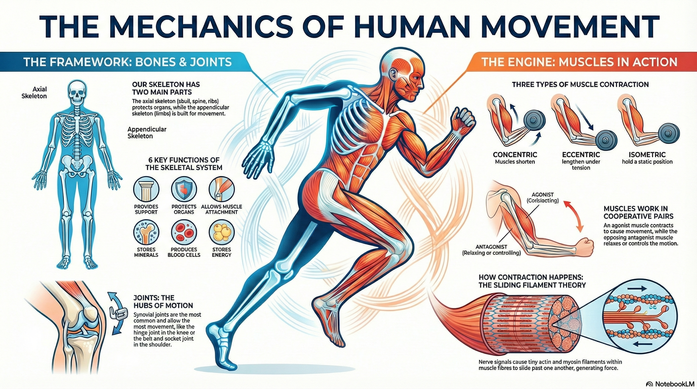
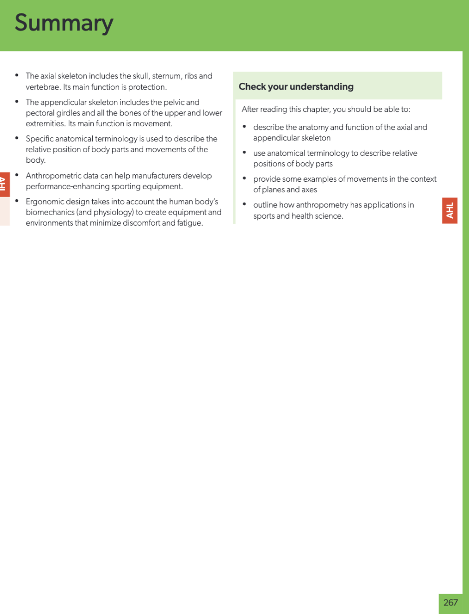
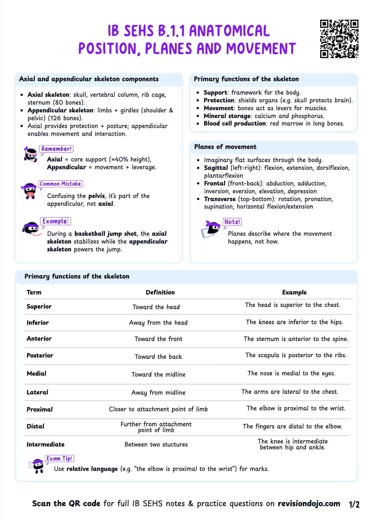
- Skeletal Divisions: The human skeleton is divided into two primary components. The axial skeleton forms the main axis of the body (skull, spine, ribs) and its primary function is protection and support. The appendicular skeleton consists of the limbs and girdles, with a primary function of movement.
- Anatomical Descriptors: A set of terms is used to describe the relative
position of
body parts:
- Superior/Inferior: Towards the head / towards the feet.
- Anterior/Posterior: Towards the front / towards the back.
- Medial/Lateral: Towards the midline / away from the midline.
- Proximal/Distal: Closer to the trunk / further from the trunk (used for limbs).
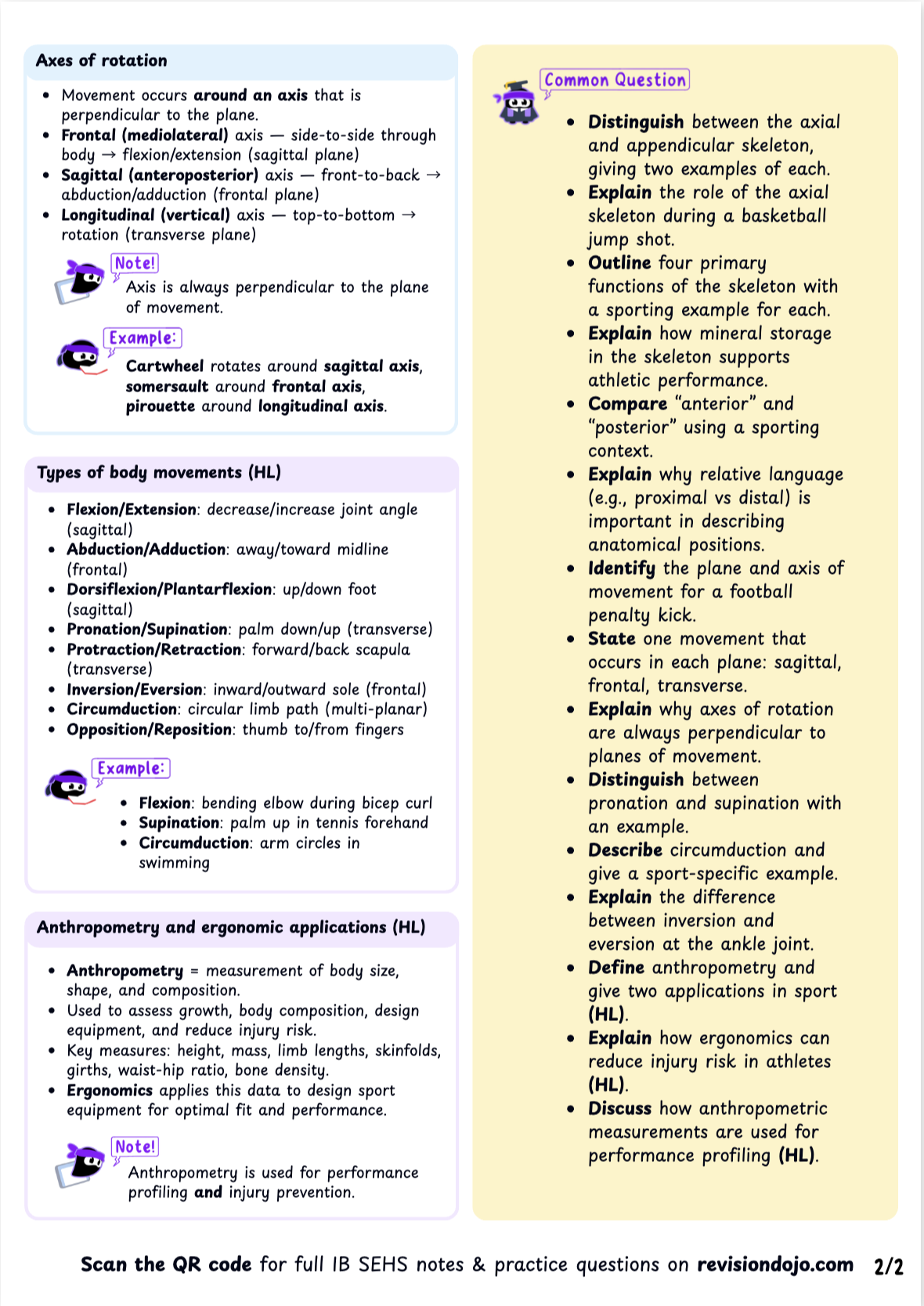
- Planes and Axes: Movement occurs in a plane and rotates around an axis.
These
pairings are fundamental to biomechanical analysis:
- Movement in the sagittal plane (e.g., flexion, extension) occurs around a frontal axis. This plane divides the body into left and right halves.
- Movement in the frontal plane (e.g., abduction, adduction) occurs around a sagittal axis. This plane divides the body into front and back halves.
- Movement in the transverse plane (e.g., rotation) occurs around a longitudinal (or vertical) axis. This plane divides the body into upper and lower halves.
- Common movements include flexion, extension, adduction, abduction, rotation, pronation, supination, plantarflexion, and dorsiflexion.
A tennis coach can use this precise language to improve a player's serve. They might instruct the player to increase shoulder abduction (frontal plane) and external rotation (transverse plane) during the backswing to generate more power. This is more effective than vague feedback like "bring your arm back more," as it provides a clear, biomechanically correct instruction that can be analyzed and replicated.
[INSERT FIGURE: Diagram of the skeletal system and diagram of the major planes found in the SEHS data booklet]
Understanding the skeletal framework is the first step; next, we must examine the connective tissues and joints that hold this framework together and enable it to move.
B.1.2 Structure and function of connective tissues and joints
The intricate interaction between stability and mobility in the human body is governed by the properties of its connective tissues and the specific design of its joints. Some joints are built for maximum stability at the expense of motion, while others sacrifice stability to allow for a wide range of movement, each tailored to its unique functional demands.
Overview This sub-section explores the passive structures that permit and limit movement. It covers the roles of key connective tissues like ligaments and tendons and classifies the three main types of joints (articulations) based on their structure and the degree of movement they allow.
Core Concepts
- Connective Tissues: Various tissues connect, support, and bind other
body tissues.
Key examples in movement include:
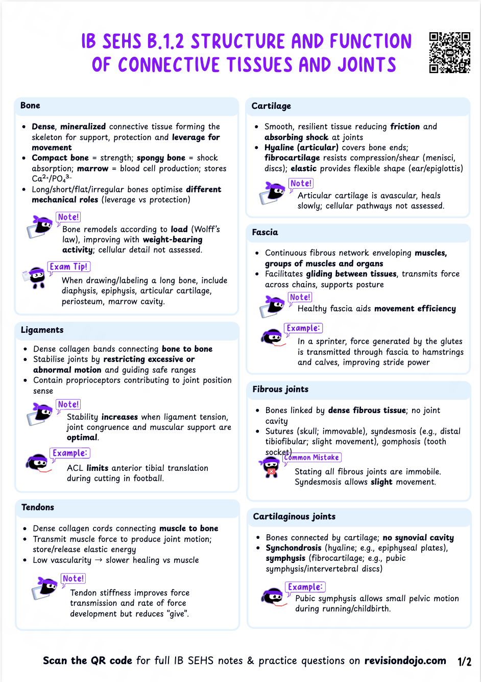
- Bone: Provides the rigid framework for the body.
- Cartilage: A smooth, elastic tissue that reduces friction and absorbs shock in joints.
- Ligaments: Connect bone to bone, providing stability to joints.
- Tendons: Connect muscle to bone, transmitting the force of muscle contraction.
- Fascia: A sheet of connective tissue that encloses muscles and other organs.
- Joint Classifications: Joints are classified by their structure and the
movement
they permit:
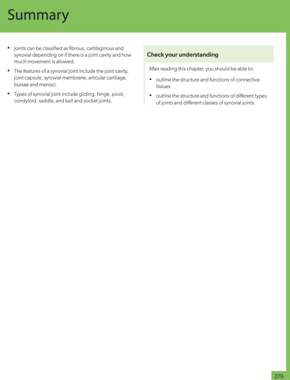
- Fibrous joints (e.g., skull sutures) are immovable and offer maximum stability.
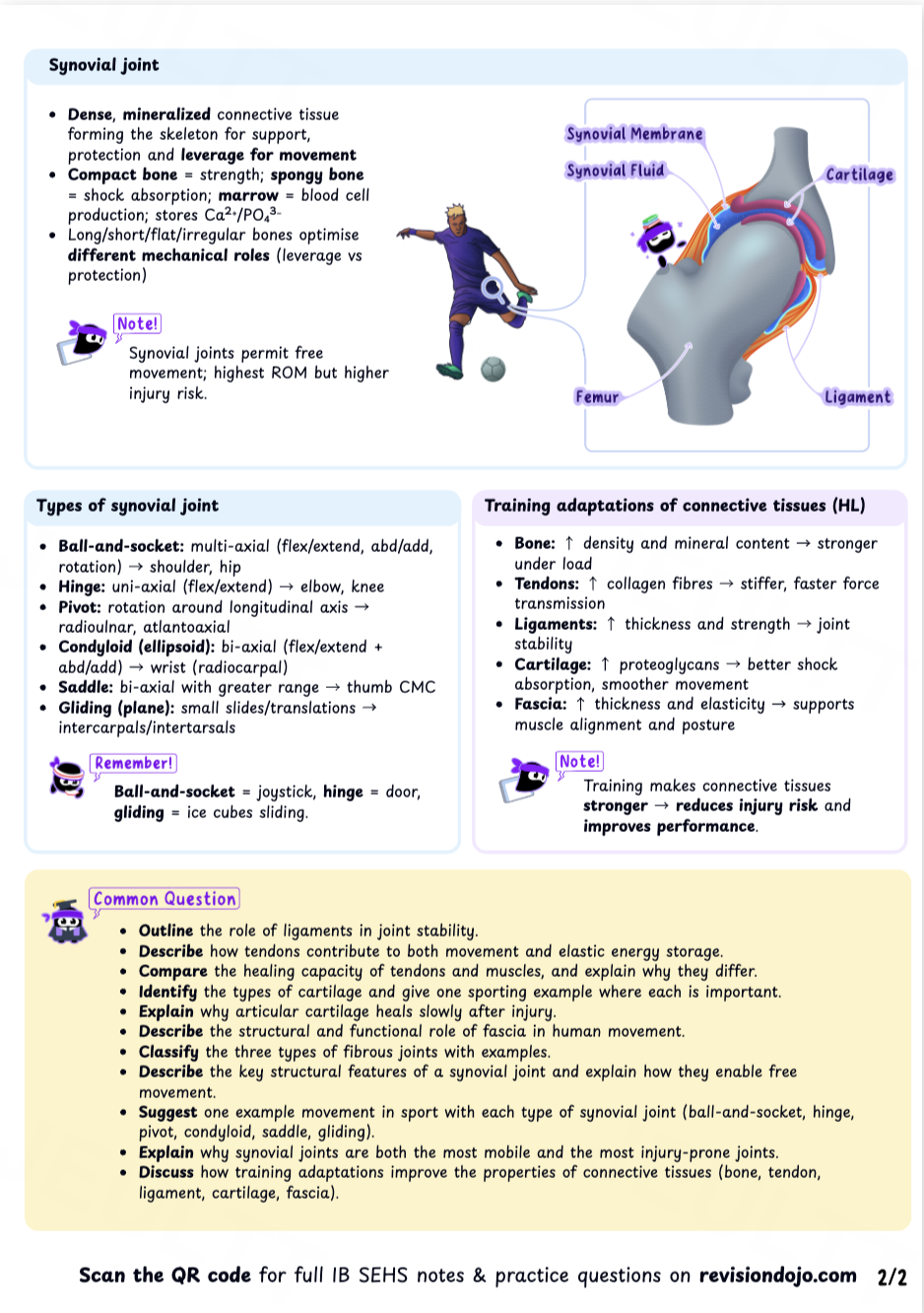
- Cartilaginous joints (e.g., between vertebrae) allow for limited movement.
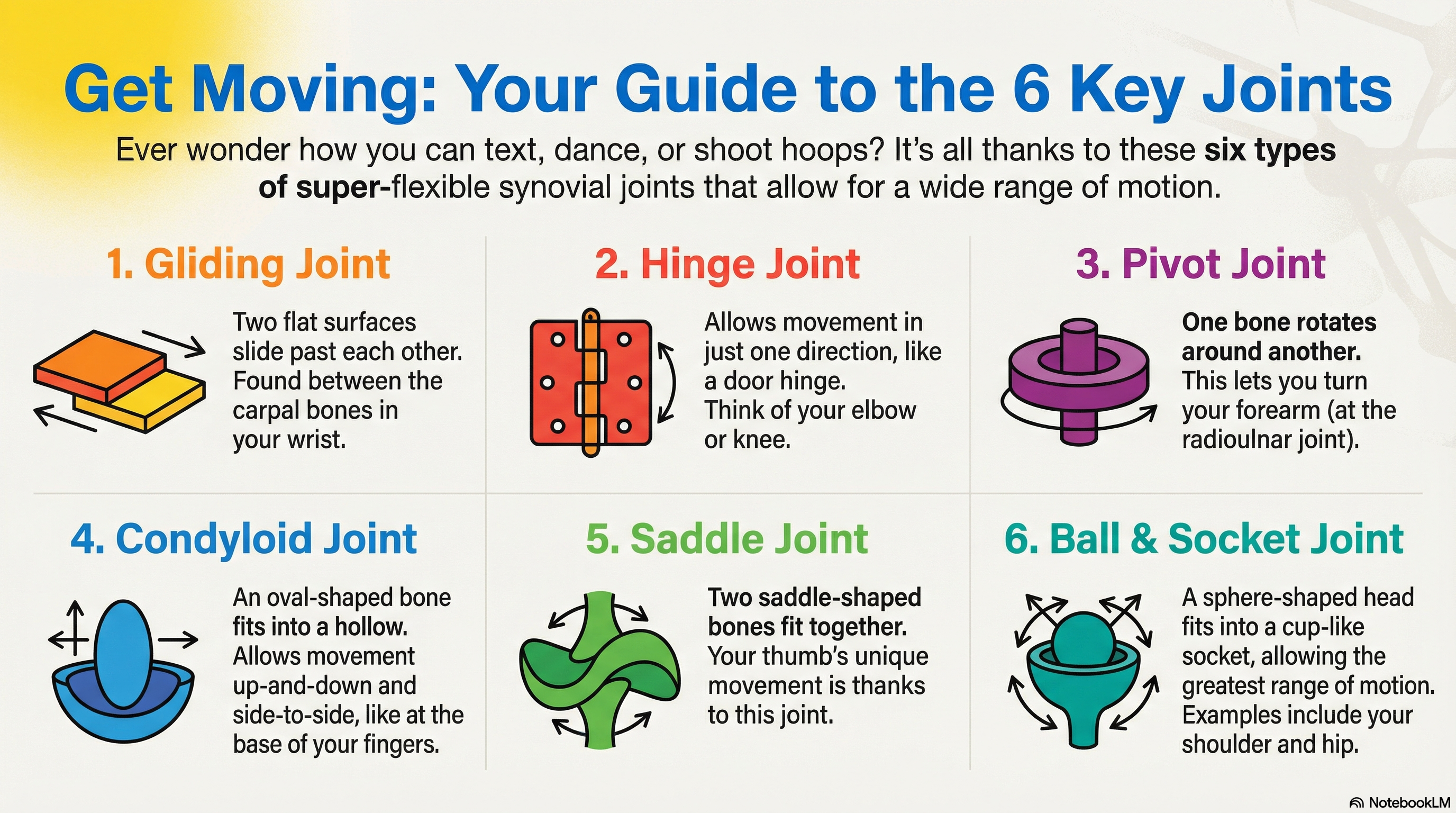
- Synovial joints (e.g., knee, shoulder) are freely movable and are the most common type of joint in the body. Different classes of synovial joints, such as hinge or ball-and-socket, provide varying degrees of stability and allow for different types of movement.
A baseball pitcher exemplifies the trade-off between mobility and stability. The shoulder is a synovial ball-and-socket joint, allowing for extreme rotation and circumduction, which is essential for throwing a ball at high velocity. However, this high mobility means it has less inherent stability compared to a joint like the knee. It relies heavily on ligaments and muscles for support, making it highly susceptible to dislocations and overuse injuries.
These passive joint structures provide the potential for movement, but it is the active contraction of muscles that generates the force to create it.
B.1.3 Muscular function
Understanding muscular contractions is of strategic importance as they are the "engine" of all human movement. From the microscopic interaction of protein filaments at the cellular level to the complex, coordinated actions required for elite athletic performance, muscles are the active force generators that move the skeletal system.
Overview This section focuses on how muscles function to produce force and movement. It introduces the concept of the motor unit, explains the different types of muscle contractions, and for HL students, delves into the molecular mechanism of muscle action known as the sliding filament theory.
Core Concepts
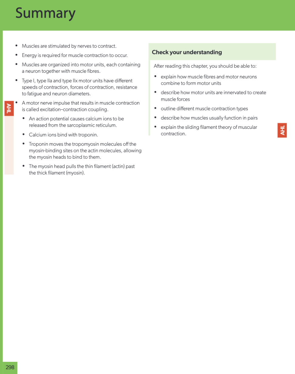
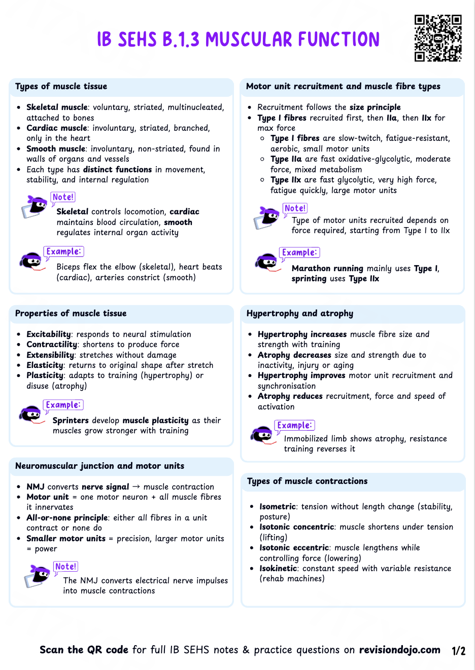
- Motor Units and Recruitment: A motor unit consists of a single motor neuron and all the muscle fibers it innervates. When a motor unit is activated, it follows the all-or-none principle—all fibers in that unit contract maximally, or not at all. The body recruits different fiber types based on the demands of the activity: slow-twitch Type I fibers for endurance, and fast-twitch Type IIa and Type IIx fibers for powerful, explosive movements.
- Types of Contractions:
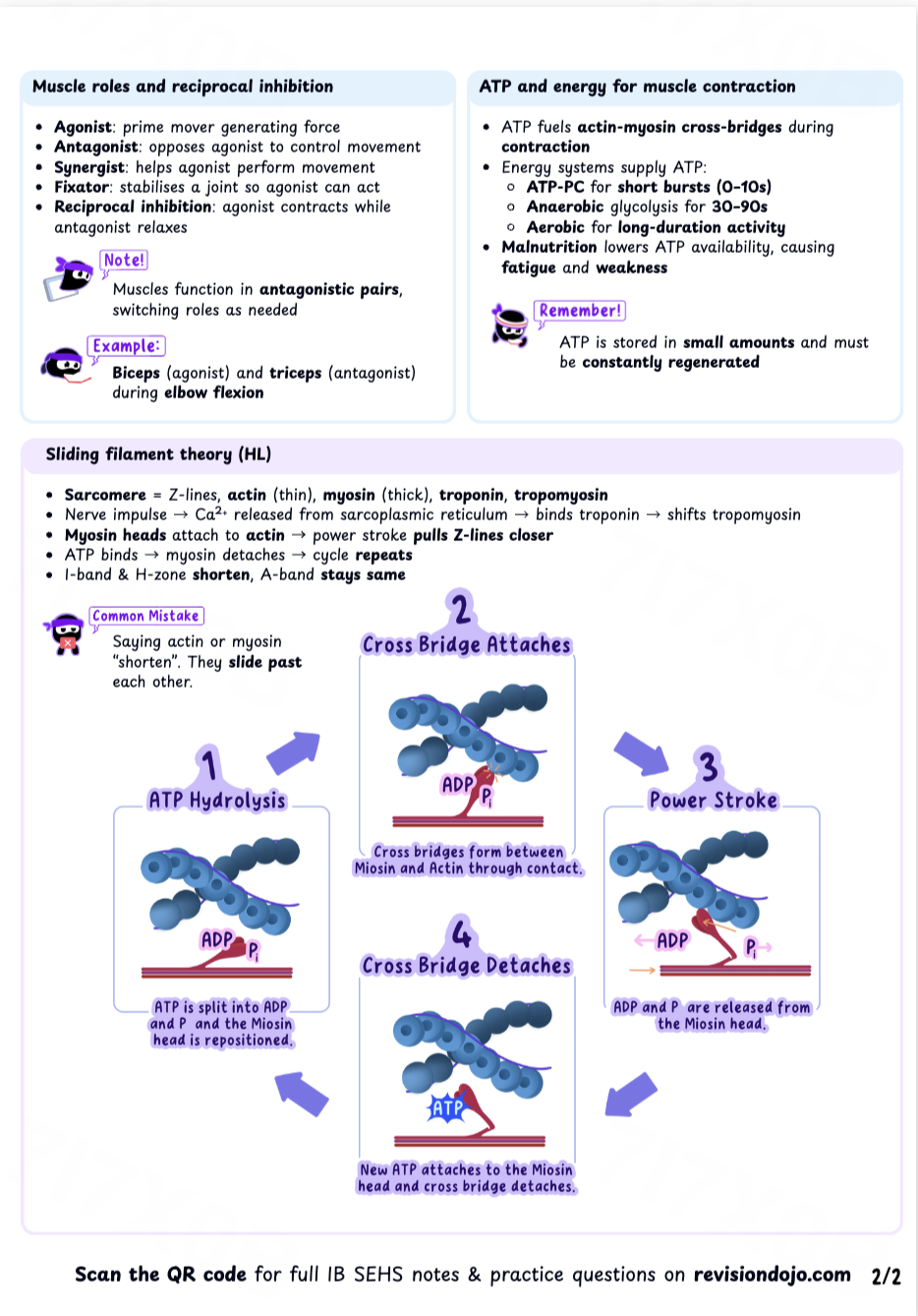
- Isometric: Muscle generates force without changing length (e.g., holding a plank).
- Isotonic Concentric: Muscle generates force while shortening (e.g., the upward phase of a bicep curl).
- Isotonic Eccentric: Muscle generates force while lengthening (e.g., lowering a weight slowly in a bicep curl).
- Sliding Filament Theory (HL Only): This theory explains contraction at the microscopic level. It involves the roles of calcium ions, which bind to troponin, causing tropomyosin to move and expose binding sites on the actin filament. The myosin heads then attach to actin, using energy from ATP to pull the actin filaments closer together, shortening the sarcomere.
A weightlifter performing a bench press utilizes a combination of muscle contractions. The upward push of the barbell is an isotonic concentric contraction of the pectoral and triceps muscles. Lowering the bar back to the chest in a controlled manner is an isotonic eccentric contraction. This controlled eccentric phase is crucial for building muscle strength and hypertrophy, as it places significant tension on the muscle fibers.
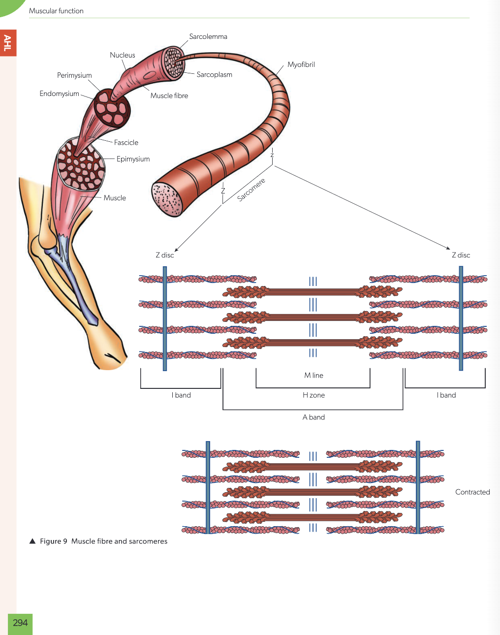
Diagrams of a sarcomere and a muscle fibre (SEHS data booklet)
The force generated by muscles is applied to the skeleton, which functions as a system of levers to create movement.
B.1.4 Levers in movement and sport
The principles of mechanical levers, fundamental to physics and engineering, can be directly applied to the human musculoskeletal system and to sports equipment. Analyzing the body as a system of levers allows us to understand and optimize force production, speed of movement, and overall efficiency.
Overview This sub-section applies mechanical principles to human movement. It introduces the three classes of levers, explains how their structure provides either a mechanical advantage or disadvantage, and provides examples from within the body and in sporting equipment.
Core Concepts
- Classes of Levers: Levers are classified based on the relative
positions of the
fulcrum (pivot point), effort (muscle force), and load (resistance).
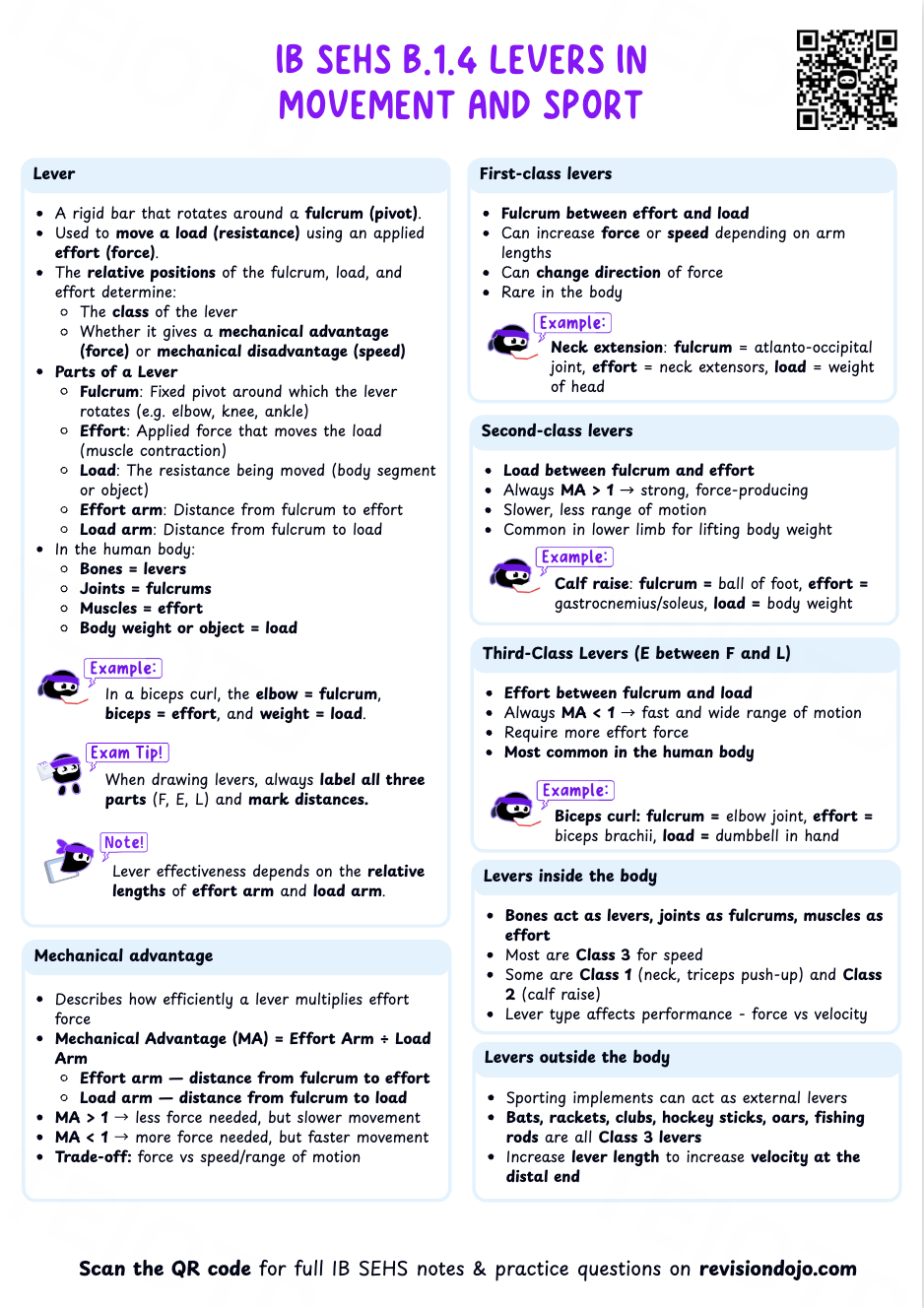
- First-Class: Fulcrum is between the effort and the load (e.g., a seesaw).
- Second-Class: Load is between the fulcrum and the effort (e.g., a wheelbarrow).
- Third-Class: Effort is between the fulcrum and the load (e.g., tweezers).
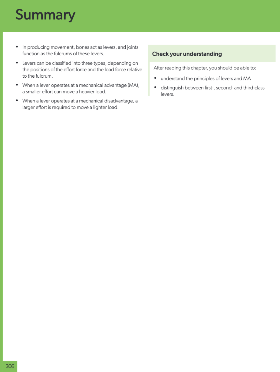
- Mechanical Advantage: The arrangement of a lever determines its function. Second-class levers provide a mechanical advantage, meaning the force produced is greater than the effort applied, making them effective for moving heavy loads. Third-class levers, the most common in the human body, operate at a mechanical disadvantage but provide a large range of motion and high speed at the end of the lever, which is ideal for athletic movements.
First-Class Lever: Extending the neck to look upwards, where the atlanto-occipital joint is the fulcrum, neck muscles provide the effort, and the head is the load.
Second-Class Lever: Performing a calf raise (plantarflexion), where the ball of the foot is the fulcrum, the calf muscles provide the effort, and the body's weight is the load.
Third-Class Lever: A bicep curl, where the elbow joint is the fulcrum, the biceps muscle insertion is the effort, and the weight in the hand is the load.
External Lever: A tennis player with a longer racquet can increase the velocity of the racquet head when serving. While it may require more effort to swing, the longer lever arm amplifies the speed at the point of impact, resulting in a faster serve.
Applying this theoretical knowledge is the final step, best accomplished through practice with exam-style questions.
Why A is wrong: Abduction is a sideways movement away from the midline, occurring in the frontal plane.
Why B is wrong: Rotation of the head occurs in the transverse plane, around a vertical axis.
Why D is wrong: Lateral flexion (bending the spine to the side) occurs in the frontal plane.
Why A is wrong: This contradicts the "all" part of the principle.
Why C is wrong: The strength of contraction for a single motor unit is maximal; the overall muscle force is graded by recruiting more motor units, not by varying the contraction strength of one unit.
Why D is wrong: The all-or-none principle refers to the activation of a motor unit, not the type of overall muscle contraction (isometric vs. isotonic).
Why A is wrong: Elbow extension is an example of a first-class lever.
Why B is wrong: A calf raise is a classic example of a second-class lever.
Why D is wrong: Nodding the head is an example of a first-class lever.
Why A is wrong: ATP provides the energy for the power stroke.
Why B is wrong: The binding of a new ATP molecule to the myosin head causes it to detach from actin.
Why D is wrong: The myosin heads pull the actin filaments; calcium is the trigger, not the transporter.
The questions below provide insight into the types of challenges you will face in Paper 1B. Unlike standard knowledge checks, this component places a distinct emphasis on data analysis and experimental work.
Success in Paper 1B requires you to apply the "Nature of Science" (NOS) skills—such as evaluating methodologies, interpreting graphs, and understanding study design—rather than simply recalling course content.
To access a complete archive of true past papers and exemplar materials for Paper 1B, please use the resource link below.
The provided source document (SEHS Syllabus Guide) does not contain specific data sets, graphs, or tables related to topic B.1 that would allow for the creation of a data-based question as per the directive. Therefore, this section cannot be completed without violating the 'No Therefore, this section cannot be completed without violating the 'No Hallucination' directive.
No mathematical formulas or specific calculations are identified within syllabus topic B.1 Generating movement in the body.
Certain concepts in biomechanics are often confused due to their nuanced relationship. Clarifying these common points of confusion is key to developing a robust and accurate understanding of how the body moves.
IB SEHS is an integrated course where concepts from different topics are interconnected. Being able to make connections between topics is a high-level skill that demonstrates a deep conceptual understanding. The syllabus guide explicitly provides "linking questions" to encourage this type of thinking.
Use the following checklist to self-assess your confidence with the core concepts in this topic. This is a tool to help you identify areas where you feel strong and areas that require further review. Go through each statement and reflect on your ability to perform the described task.
- I can distinguish between the axial and appendicular skeleton.
- I can identify the major planes of movement and provide examples of motion in each.
- I can describe the structure of a synovial joint.
- I can explain the difference between isometric, concentric, and eccentric muscle contractions.
- I can describe the all-or-none principle of motor unit contraction.
- I can identify the three classes of levers and provide an anatomical example of each.
- (HL Only) I can explain the key steps of the sliding filament theory, including the roles of ATP, calcium, actin, myosin, troponin, and tropomyosin.
🎉 Topic B.1 Mastered!
You've completed the study guide for Generating Movement in the Body. Keep practicing with exam-style questions to solidify your understanding!